💡 Direct Answer & Executive Summary (Hematocytometer Chamber Cell Density Calculator)
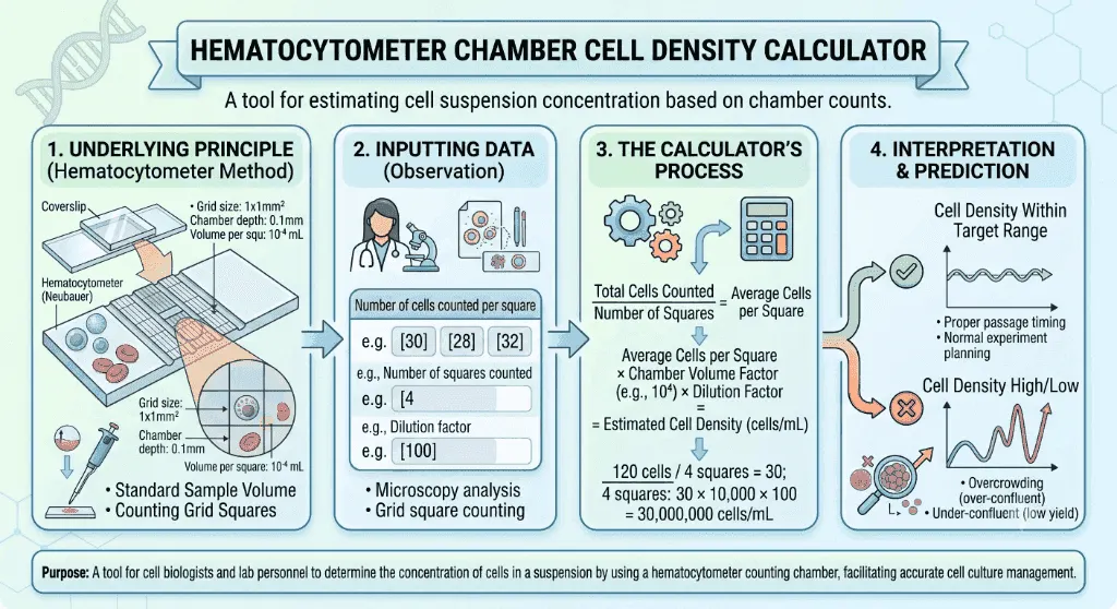
Definition: Calculate cell suspension concentration (cells/mL), total culture yield, and Trypan Blue viability percentage using a standard Neubauer hemocytometer counting chamber.
Governing Math Formula: Cell Concentration = (Total Cells Counted / Number of Large Squares) × 10,000 × Dilution Factor (DF). Total Cells in Flask = Concentration × Total Suspension Volume (mL).
Target Applications: Provides real-time quantitative solutions in Biology for students, engineers, researchers, and finance professionals.
Hematocytometer Chamber Cell Density Calculator: Quantification & Viability Guide
1. Introduction
In modern biomedical research and industrial biomanufacturing, knowing the exact concentration of living cells in a suspension is essential. When administering CAR-T cell immunotherapies, oncologists must deliver an exact therapeutic dose of of patient body weight. In pharmaceutical bioreactors, technicians culture Chinese Hamster Ovary () cells at tightly controlled seeding densities to maximize monoclonal antibody production. In clinical hematology and neurology, emergency CSF cell counts differentiate life-threatening bacterial meningitis from viral infections within minutes.
How do scientists accurately count microscopic cells suspended in liquid volumes? How do we calculate cell viability to ensure our experimental cultures are thriving rather than dying?
The gold-standard laboratory instrument that has powered quantitative cell biology for over a century is the Neubauer Hemocytometer (also spelled haemocytometer or hematocytometer).
flowchart LR
    SUSP["🧪 Cell Suspension Sample
(e.g., Trypsinized T-Flask Culture)"] --> DILUTE["🎨 Trypan Blue Staining
1:1 Mix (Dilution Factor DF = 2)"]
    DILUTE --> LOAD["🧫 Load Neubauer Chamber
Capillary Loading under Coverslip (10 µL)"]
    LOAD --> COUNT["🔬 Count 4 Large Corner Squares
Apply Top & Left Inclusion Rule"]
    COUNT --> CALC["🧮 Concentration Formula
C = (Total / Squares) × 10⁴ × DF"]
2. Definitions
2.1 Simple Everyday Definition
A Hemocytometer is a precision glass microscope slide etched with a microscopic grid of known dimensions. By counting the number of cells inside a fixed grid volume under a microscope, you can calculate the exact concentration of cells in your entire bottle or flask (measured in cells per milliliter, ).
2.2 Formal Technical Definition
A hemocytometer is a specialized volumetric counting chamber manufactured with optical tolerance tolerances. A precision ground coverslip rests on two elevated lateral glass ridges, establishing a calibrated fluid depth of exactly () over a precision-etched grid.
The concentration of cells in the original stock suspension () is given by:
Where:
- is the total number of cells counted across large squares.
- ().
- The multiplication factor is .
- is the volumetric Dilution Factor (e.g., for a $1:1$ cell-to-dye mixture).
2.3 Vivid Real-World Analogies
The City Block Population Grid:
Imagine trying to count 5 million people living across a vast metropolitan area. Instead of counting every individual street, you select 4 standard 1-square-kilometer city blocks, count the residents inside them, calculate the average density per block, and multiply by the total city area. The hemocytometer does this on a microscopic scale.
The Swimming Pool Sampling Bucket:
If you want to know how many microscopic algae cells are in a 50,000-liter pool, you dip a calibrated 100-nanoliter bucket, count the algae inside, and multiply by the ratio between the bucket volume and one liter ().
3. History & Scientific Milestones
The invention of the hemocytometer transformed medicine from qualitative observation into quantitative cellular science.
timeline
    title Evolution of Hemocytometry & Cell Counting
    1874 : Louis-Charles Malassez : Invents first capillary counting chamber at Collège de France
    1878 : Georges Hayem & Karl Bürker : Introduce improved optical grid line rulings
    1904 : Paul Ehrlich : Discovers Trypan Blue dye exclusion for viable cell staining
    1920 : Otto Neubauer : Standardizes the 'Improved Neubauer' 9-square grid system
    1953 : Wallace Coulter : Patents electronic impedance cell counter (Coulter Counter)
2010s : Digital Automated Counters : Introduce AI computer vision image-based hemocytometry- Louis-Charles Malassez (1874): French physician and physiologist who built the first hemocytometer to diagnose severe anemia by quantifying erythrocyte numbers in human blood.
- Otto Neubauer (1900s): Standardized the Improved Neubauer Grid, featuring a central grid partitioned into 25 groups of 16 mini-squares and 4 corner squares bordered by triple ruling lines, which remains the global standard ISO specification today.
- Paul Ehrlich (1904): Discovered that synthetic azo dyes like Trypan Blue selectively enter and stain dead cells with compromised plasma membranes while being actively excluded by live, metabolically intact cells.
4. Core Concepts & Theoretical Principles
graph TD
    subgraph Grid_Layout["Improved Neubauer Chamber (3mm × 3mm Grid)"]
        W1["Corner Square 1 (1 mm²)
Mammalian / WBCs"] --- T["Top Middle Square"] --- W2["Corner Square 2 (1 mm²)
Mammalian / WBCs"]
        L["Left Middle Square"] --- C["Center Square (1 mm²)
25 Groups × 16 Small Squares
RBCs / Yeast / Platelets"] --- R["Right Middle Square"]
        W3["Corner Square 3 (1 mm²)
Mammalian / WBCs"] --- B["Bottom Middle Square"] --- W4["Corner Square 4 (1 mm²)
Mammalian / WBCs"]
    end
4.1 Standard Chamber Dimensions & Volumes
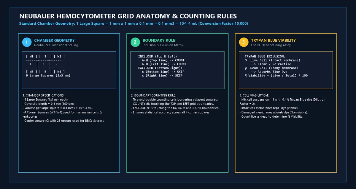
- Total Etched Grid Area: , subdivided into 9 large primary squares ( each).
- Chamber Fluid Depth: Exactly ().
- Volume of 1 Large Corner Square:
- Volume of Entire 9-Square Grid:
4.2 Which Squares to Count?
- Mammalian Cell Lines (HeLa, HEK293, T-cells, Stem Cells): Count the 4 large corner squares (). Each square is , subdivided into 16 smaller squares to guide the eye.
- Red Blood Cells (RBCs) & Brewing Yeast: Because yeast and RBCs are much smaller () and present at much higher densities (), count the 5 sub-squares inside the central square (the 4 corner sub-squares and 1 center sub-square).
4.3 The Inclusion / Exclusion Boundary Rule
To prevent counting the same cell twice across adjacent squares, microscopists adhere to the universal "Top and Left" rule (or "Bottom and Right" rule):
- COUNT: Any cell touching or overlapping the Top or Left boundary lines of the square.
- DO NOT COUNT: Any cell touching the Bottom or Right boundary lines.
5. Mathematical Formulas & Derivations
5.1 Cell Concentration Formula ()
Where:
- is the number of large squares counted (typically ).
- $10,000$ (or ) is the chamber conversion factor ().
- is the dilution factor ().
5.2 Total Yield in Suspension ()
Where is the total liquid volume of your cell suspension in milliliters ().
5.3 Percentage Cell Viability
5.4 Subculture Seeding Volume Formula ()
To determine how much cell suspension volume to pipette into a new culture vessel:
Where:
- is the required number of cells for the new flask (e.g., ).
- is the viable cell concentration ().
5.5 Variable Reference Table
| Parameter | Symbol | Units | Typical Experimental Value | Biological Role |
|---|---|---|---|---|
| Live Cells Count | Cells | Primary numerator for viable density | ||
| Dead Cells Count | Cells | Trypan blue-positive non-viable cells | ||
| Squares Counted | Squares | Denominator for mean square density | ||
| Dilution Factor | Dimensionless | Accounts for volumetric pre-dilution | ||
| Chamber Conversion Factor | ||||
| Cell Concentration | Final suspension concentration | |||
| Stock Flask Volume | Total volume in harvest tube or bioreactor | |||
| Total Viable Yield | Cells | Absolute available cell quantity |
6. Step-by-Step Computational Walkthrough
Let us calculate the cell density, viability, total yield, and passaging volume for a confluent flask of human embryonic kidney () cells:
flowchart TD
    STEP1["Step 1: Record Microscopy Raw Data
Live = 160 cells | Dead = 16 cells in 4 squares | DF = 2 | V_stock = 12 mL"] --> STEP2["Step 2: Calculate Average Cells Per Square
Avg Live = 160 / 4 = 40.0 cells/square"]
    STEP2 --> STEP3["Step 3: Calculate Concentration (cells/mL)
C = 40.0 × 10,000 × 2 = 800,000 cells/mL (8.0 × 10⁵ cells/mL)"]
    STEP3 --> STEP4["Step 4: Calculate Culture Viability
% Viability = [160 / (160 + 16)] × 100 = 90.9%"]
    STEP4 --> STEP5["Step 5: Calculate Total Yield & Seeding Volume
Total Yield = 8.0 × 10⁵ × 12 mL = 9.6 × 10⁶ viable cells
Seeding 2.0 × 10⁶ cells requires V_seed = 2.50 mL"]- Step 1: Input Observations:- Viable (clear) cells in 4 corner squares: .- Non-viable (blue) cells in 4 corner squares: .- Dilution: $1:1$ Trypan Blue dye mix ().- Total stock volume (): .- Desired seeding target for new flask: .
- Step 2: Calculate Mean Cells Per Large Square:
- Step 3: Calculate Viable Cell Concentration ():
- Step 4: Determine Culture Viability Percentage:
- Step 5: Calculate Total Available Cell Yield:
- Step 6: Calculate Seeding Volume for New Flask ():(Add cell stock fresh medium to reach final volume).
7. Visual Explanations & Neubauer Grid Anatomy
graph TD
    LOAD_SAMPLE{"Prepare 10 µL Cell Suspension"}
    LOAD_SAMPLE --> COVERSLIP["Place Heavy Optical Coverslip on Rails"]
    COVERSLIP --> CAPILLARY["Introduce Pipette Tip at Notch
Capillary Action fills Chamber without Overflow"]
    CAPILLARY --> MICROSCOPE["Focus at 100x / 400x Brightfield"]
    MICROSCOPE --> DENSITY_CHECK{"Is Count Between 20 - 50 Cells/Square?"}
    DENSITY_CHECK -->|"Too Crowded (>100 cells)"| DILUTE_MORE["Dilute Sample 1:5 or 1:10 and Reload"]
    DENSITY_CHECK -->|"Too Sparse (<15 cells)"| CENTRIFUGE["Centrifuge and Resuspend in Smaller Volume"]
    DENSITY_CHECK -->|"Optimal (20-50 cells)"| COUNT_EXEC["Execute 4 Corner Counts using Top & Left Rule"]
8. Comparative Analysis & Laboratory Standards
8.1 Counting Chamber Model Specifications
| Chamber Type | Grid Dimensions | Chamber Depth | Volume per Large Square | Primary Recommended Use |
|---|---|---|---|---|
| Improved Neubauer | () | Universal (Mammalian cells, WBCs, yeast, RBCs) | ||
| Fuchs-Rosenthal | () | total | Low-density fluids (CSF cell counts, urine sediment) | |
| Malassez | () | Hematology cell counting (Popular in Europe) | ||
| Nageotte | Ultra-low density residual WBCs in leukoreduced blood | |||
| Thoma | () | High-density suspensions (RBCs, spermatozoa, yeast) |
8.2 Standard Culture Vessel Seeding Densities
| Vessel Type | Growth Surface Area | Recommended Seeding Cell Count | Standard Working Media Volume |
|---|---|---|---|
| 96-Well Plate | |||
| 24-Well Plate | |||
| 6-Well Plate | |||
| T-25 Flask | |||
| T-75 Flask | |||
| T-175 Flask |
9. Practical Real-World Applications
Example 1: Passaging CHO Cells in Monoclonal Antibody Production
A bioprocess engineer harvests a suspension CHO-K1 cell culture from an orbital shake flask.
- Counts in 4 Large Squares: ($1:1$ Trypan blue dilution, ).
- Viable Concentration:
- Viability: .
- Seeding Target: Inoculating into a new bioreactor requires pipetting .
Example 2: Emergency Diagnostic Lumbar Puncture (CSF Pleocytosis)
A patient with high fever, neck stiffness, and confusion undergoes an emergency lumbar puncture.
- Undiluted CSF Loaded into Neubauer Chamber ():
- Across 4 large corner squares, the technician counts .
- WBC Concentration:
- Normal adult CSF contains . This massive pleocytosis confirms acute bacterial meningitis, prompting immediate intravenous cephalosporin therapy.
Example 3: Brewing Yeast Pitching Rate Calculation
A master brewer pitches lager yeast (Saccharomyces pastorianus) into a batch of wort at .
- Required pitch rate .
- Total yeast cells required .
- Hemocytometer slurry count . Slurry volume needed .
Example 4: Normalizing Cell Inputs for MTT Cytotoxicity Assays
In drug screening, researchers seed exactly per well in a 96-well plate ( volume).
- Required working concentration .
- Using the hemocytometer density of the harvested stock (), the stock is diluted with complete growth medium before plating.
10. In-Depth Case Studies
Case Study 1: Industrial CHO-K1 Bioreactor Seeding & Passaging
- Background: In commercial biopharmaceutical manufacturing, maintaining consistent seeding density is critical for glycosylation quality and antibody titer.
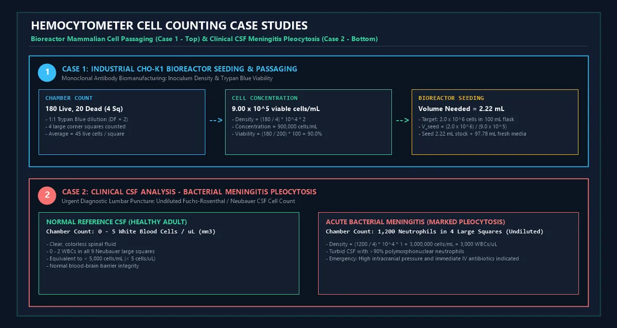
- Harvest & Chamber Count: A seed train flask is harvested. A $1:1$ Trypan Blue dilution () is loaded into an Improved Neubauer chamber:- 4 Large Corner Squares: (Total ) and .
- Calculations:- Viable Concentration: .- Viability: (Meets the threshold required for subculturing).- Total Stock Yield: .
- Inoculation Execution: Seeding a new production flask at requires . The technician pipettes of stock culture.
Case Study 2: Clinical CSF Analysis in Acute Bacterial Meningitis
- Background: A 19-year-old college student presents to the Emergency Department with severe headache, photophobia, nuchal rigidity, and petechial rash.
- Lumbar Puncture: Cerebrospinal fluid appears cloudy/turbid. An aliquot of uncentrifuged, undiluted CSF () is loaded immediately into a Neubauer hemocytometer.
- Laboratory Count:- Corner Squares 1 to 4: (Total ).
- Calculations:
- Clinical Significance: Identifies severe neutrophilic pleocytosis (), triggering urgent isolation and intravenous ceftriaxone + vancomycin administration, saving the patient's life.
11. Advantages of Hemocytometer Chamber Counting
- Direct Gold Standard Reference: Unaffected by electronic calibration drifts, optical autofluorescence, or cell shape anomalies that can mislead automated counters.
- Simultaneous Viability Assessment: Direct visual distinction of membrane integrity via Trypan Blue exclusion dye.
- Extreme Cost Efficiency: A single high-grade quartz hemocytometer can be cleaned, autoclaved, and reused for decades with zero consumable cartridge costs.
- Immediate Qualitative Feedback: Allows simultaneous visual inspection for bacterial/fungal contamination, cell clumping, and morphologic apoptosis.
12. Methodological Complexities & Artifacts
graph LR
    subgraph Artifacts["Hemocytometry Errors & Artifacts"]
        E1["Chamber Overfilling / Flooding
Floating coverslip increases volume!"]
        E2["Cell Clumping / Aggregation
Incomplete enzymatic detachment"]
        E3["Trypan Blue Toxicity
Incubation > 10 min kills live cells"]
        E4["Boundary Bias
Inconsistent top/bottom counting"]
    end
Artifacts -->|"Results In"| ERR["⚠️ Inaccurate Concentration & Seeding Failure"]- Coverslip Floating (Overfilling): If excess liquid overflows into the lateral V-trenches, surface tension lifts the coverslip, increasing the chamber depth beyond and falsely inflating cell counts by .
- Trypan Blue Cytotoxicity: Trypan Blue is cytotoxic. Cells left in dye solution for more than 10 minutes begin taking up dye non-specifically, producing falsely low viability readings.
- Incomplete Trypsinization & Clumping: Clusters of 3+ cells must be thoroughly triturated before loading. If clumping persists, counts become statistically invalid.
13. Common Mistakes to Avoid
1. Forgetting to Multiply by the Dilution Factor ():
When performing a $1:1$ Trypan Blue viability assay, you have diluted your sample . Omitting the multiplier halves your true cell concentration!
2. Counting Fewer than 100 Total Cells:
Counting fewer than 100 total cells across the 4 corner squares introduces substantial Poisson sampling variance ( error). If cells are sparse, count all 9 large squares or concentrate the suspension.
3. Using a Standard Thin Glass Coverslip (No. 1.5):
Standard microscopy coverslips are too flexible and bow under surface tension. Always use a heavy ground optical glass coverslip ( thickness) designed specifically for hemocytometers to ensure exact depth.
12. Frequently Asked Questions (FAQ)
What is the standard volume of one large corner square in a Neubauer hemocytometer?
The volume of one large square () at a standard depth of is:
Why do we multiply by $10,000$ (or ) in the formula?
Because , one large square () represents of a milliliter. Multiplying by $10,000$ converts the count in one square into the number of cells per milliliter ().
How is cell viability calculated using Trypan Blue?
Live cells with intact membranes exclude the dye, while dead cells stain dark blue.
What is the boundary inclusion/exclusion rule?
To avoid double counting, count all cells touching the Top and Left boundary lines of a square, and exclude any cells touching the Bottom and Right boundary lines.
What is the ideal cell concentration for hemocytometer counting?
The optimal counting range is (corresponding to in the loaded chamber).
What if my cell suspension is too dense (>100 cells/square)?
Perform a serial dilution (e.g., $1:5$ or $1:10$) in PBS or growth media before mixing with Trypan Blue, and multiply by the combined dilution factor.
What if my cell suspension is too dilute (<15 cells/square)?
Centrifuge your harvest tube (e.g., for 5 minutes), aspirate a portion of the supernatant, and resuspend the cell pellet in a smaller volume (e.g., instead of ) before recounting.
15. Expert Tips for Cell Biologists & Technicians
- Verify Newton's Refraction Rings: When pressing the heavy coverslip onto the moistened chamber rails, look for iridescent rainbow interference bands (Newton's rings), confirming proper physical contact and exact depth.
- Mix Thoroughly Immediately Before Loading: Cells settle rapidly in pipette tips. Always vortex or gently invert your cell-dye mixture immediately before loading the chamber.
- Load by Pure Capillary Action: Place the pipette tip at the notch where the coverslip meets the glass floor and release . Allow capillary force to pull the liquid smoothly across the grid without pushing air bubbles or flooding trenches.
- Clean with 70% Ethanol and Lint-Free Lens Paper: Never clean hemocytometers with coarse paper towels, which scratch precision grid etchings.
16. Summary Checklist
- ✔ Mount Coverslip: Ensure Newton's interference rings are visible on glass support rails.
- ✔ Prepare Sample: Mix cell suspension $1:1$ with 0.4% Trypan Blue ().
- ✔ Load Chamber: Introduce per side; verify absence of bubbles or flooding.
- ✔ Microscopic Survey: Focus under objective ( total magnification).
- ✔ Count 4 Corner Squares: Record live and dead counts using the Top-and-Left boundary rule.
- ✔ Calculate Concentration: Apply .
- ✔ Calculate Viability & Yield: Determine Viability and total available cells ().
Additional Technical Guidelines & Measurement Standards
When conducting calculations for Hematocytometer Chamber Cell Density Calculator, maintaining quantitative precision and verifying input parameter boundaries is essential for reliable scenario evaluation. Always verify that raw numerical inputs are measured using standardized instrumentation, and double-check unit conversions prior to applying outputs in commercial, industrial, or academic projects.
For quantitative unit conversions and scientific calculations, always maintain at least four decimal places of precision during intermediate arithmetic stages before rounding final results. When preparing technical reports, laboratory documentation, or financial forecasts, cross-verify all output variables against established international measurement standards (such as SI, NIST, and ISO metric benchmarks) to guarantee consistent analytical rigor across all computational workflows.
MathsLover.com delivers this interactive solver 100% free of charge to foster global mathematical literacy, educational accessibility, and data-driven problem solving across scientific, technical, and academic communities worldwide.